
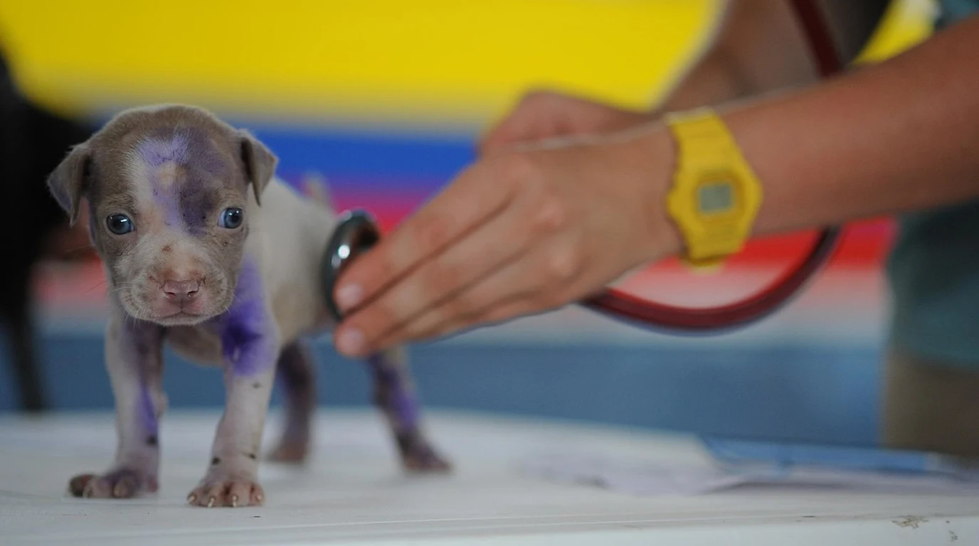
What Is a Portosystemic Liver Shunt (PSS) in Dogs?
When a portosystemic shunt is present, an anomalous vascular vessel bypasses this filtration pathway. As a consequence, neurotoxic byproducts of digestion—most notably free ammonia ($NH_3$), aromatic amino acids, short-chain fatty acids, and endogenous benzodiazepine-like compounds—are swept directly into the vena cava and systemic arterial circulation. When these toxins cross the blood-brain barrier, they trigger central nervous system edema, astrocytic swelling, and acute hepatic encephalopathy (HE).
Portosystemic shunts are categorized into two fundamental anatomical variants:
- Extrahepatic Shunts: The anomalous vessel is located outside the liver parenchyma, typically connecting the portal vein or splenic vein to the caudal vena cava or azygos vein. These account for roughly 65% to 75% of congenital cases and occur predominantly in small and toy breeds such as Yorkshire Terriers, Miniature Schnauzers, Shih Tzus, and Pugs.
- Intrahepatic Shunts: The shunt vessel traverses the liver lobes internally, frequently representing a failure of the fetal ductus venosus to close at birth. Intrahepatic shunts are significantly more common in large and giant breeds, including Irish Wolfhounds, Golden Retrievers, and Labrador Retrievers, requiring specialized interventional radiology (transvenous coil embolization).
Clinical Signs: Recognizing Hepatic Encephalopathy
Symptoms of canine liver shunts can be episodic and perplexing to pet parents. Signs frequently intensify within 30 to 90 minutes following a meal containing high-biological-value animal protein, as intestinal bacteria break down dietary amino acids into ammonia:
| Organ System | Common Clinical Manifestations | Pathophysiological Mechanism |
|---|---|---|
| Neurological (HE) | Head-pressing, aimless pacing, ataxia, blindness, seizures | Circulating ammonia crossing blood-brain barrier causing cerebral edema |
| Gastrointestinal | Intermittent vomiting, anorexia, pica, diarrhea, copper-tinged iris | Gastric ulceration secondary to impaired portal blood flow and endotoxemia |
| Urinary Tract | Stranguria, hematuria, ammonium urate bladder stones | Excessive excretion of uric acid and ammonia filtering into renal tubules |
| Somatic / Growth | Failure to thrive, stunted body mass, poor hair coat, prolonged anesthetic recovery | Inability of the liver to clear anesthetic drugs or synthesize hepatic growth factors |
Definitive Diagnostics: Pre- and Post-Prandial Bile Acids
Routine serum biochemistry often provides initial clues: low Blood Urea Nitrogen (BUN) due to impaired hepatic urea cycle conversion, low albumin (hypoalbuminemia), mild elevations in ALT and ALP, and low fasting blood glucose. However, definitive functional diagnosis requires specialized screening:
- Serum Bile Acid (SBA) Testing: The gold standard functional screen. A baseline blood sample is drawn after a 12-hour fast. The dog is then fed a small fatty meal to contract the gallbladder, and a second sample is taken exactly 2 hours later. In healthy dogs, post-prandial bile acids remain under 20 µmol/L. Dogs with portosystemic shunts frequently register levels exceeding 100 to 250 µmol/L, as escaped bile acids bypass hepatic reabsorption.
- Fasting Blood Ammonia: Elevated resting venous ammonia ($>60 ext{–}80\ \mu ext{mol/L}$) is highly suggestive of shunting or severe hepatic dysfunction, though blood samples must be analyzed immediately on ice to prevent artificial in vitro elevation.
- Abdominal Doppler Ultrasound & CT Angiography: High-resolution Doppler ultrasonography identifies anomalous vascular velocity and microhepatica (abnormally small liver). Dual-phase CT angiography serves as the definitive pre-surgical roadmap to pinpoint vessel trajectory and rule out multiple acquired shunts.
The Nutritional Protocol: Formulating a Low-Ammonia Diet
Unlike dogs in acute liver failure where protein is severely restricted, dogs with portosystemic shunts require sufficient protein to support somatic growth and prevent skeletal muscle wasting, but the protein must be sourced meticulously to minimize ammonia generation:
- Modified Protein Sourcing (Dairy and Vegetable Basis): Red meat, organ meats, and poultry contain high levels of aromatic amino acids (phenylalanine, tyrosine, tryptophan) and produce heavy ammonia loads during microbial digestion. Formulations built on cottage cheese, yogurt, eggs, and purified soy protein are vastly superior. These proteins produce less ammonia and maintain a higher ratio of branched-chain amino acids (BCAAs: leucine, isoleucine, valine), which are metabolized primarily by skeletal muscle rather than the liver.
- High-Fiber Inclusion: Fermentable soluble fibers (such as pure canned pumpkin, beet pulp, and psyllium husk) acidify intestinal lumen pH, which traps ammonia as non-absorbable ammonium ($NH_4^+$) and accelerates colonic transit time, minimizing microbial contact.
- Small, Frequent Meals: Dividing the dog’s daily caloric intake into 4 to 6 micro-meals prevents large post-prandial surges of portal toxins, keeping blood ammonia levels within a manageable neurological threshold.
Medical Stabilization Therapy: Lactulose & Antimicrobials
Prior to any surgical consultation, medical stabilization is mandatory to protect brain parenchyma:
- Lactulose Administration: A synthetic, non-absorbable disaccharide administered orally (typically 0.25 to 0.5 mL/kg q8–12h). In the colon, bacteria ferment lactulose into lactic, formic, and acetic acids. This acidification ionizes ammonia ($NH_3$) into ammonium ($NH_4^+$), which cannot permeate the intestinal mucosa and is safely expelled in soft feces. The target dose is calibrated to produce 2 to 3 soft, pudding-like stools daily.
- Gut-Targeted Antimicrobials: Low-dose oral metronidazole (7.5 mg/kg q12h) or amoxicillin is prescribed to suppress urease-producing enteric bacteria (such as Klebsiella and Proteus species), directly halving gut ammonia generation.
- Anticonvulsant Prophylaxis: In patients experiencing seizures or severe hepatic encephalopathy, levetiracetam (Keppra) is initiated at 20 mg/kg q8h. Unlike phenobarbital, levetiracetam does not undergo extensive hepatic metabolism, rendering it exceptionally safe for liver-compromised canines.
Surgical Attenuation & Post-Operative Hemodynamics
While medical and dietary therapy stabilizes the patient, definitive resolution for single congenital extrahepatic shunts requires surgical intervention by a board-certified veterinary surgeon. The historical approach of acute complete surgical ligation has been largely abandoned due to the severe risk of acute, fatal portal hypertension. Instead, modern veterinary surgery relies on gradual, progressive vascular occlusion devices:
- Ameroid Constrictor Ring: The gold standard device consisting of an inner ring of compressed casein surrounded by a stainless steel collar. When placed around the shunt vessel, the casein slowly absorbs peritoneal fluid and expands inward, while provoking a sterile localized fibrous tissue reaction that gradually occludes the vessel over a 4 to 8-week window. This gradual closure allows the hypoplastic intrahepatic portal vasculature time to expand and adapt to increased blood flow without triggering portal hypertension.
- Cellophane Banding: An alternative technique utilizing surgical cellophane folded around the shunt vessel, which incites an intense perivascular inflammatory response, slowly obliterating the vascular lumen through gradual fibrosis over several weeks.
- Post-Ligation Monitoring: Following surgery, patients are monitored intensively for signs of portal hypertension, which include acute abdominal pain, hypovolemic shock, hemorrhagic diarrhea, and rapid ascites accumulation. Medical management with lactulose and dietary protein modification must be maintained for at least 8 to 12 weeks post-operatively until follow-up bile acids testing proves full hepatic vascular remodeling and functional recovery.
When assessing canine metabolic lab panels, renal filtration and hepatic blood processing work in close concert. Review our clinical breakdown of canine liver shunt diagnostics: Canine SDMA vs Creatinine: Early Kidney Biomarkers.