
It happens in an instant: your dog sprints across the backyard chasing a ball, lets out an abrupt, high-pitched yelp, and pulls up on three legs. Within hours, they are “toe-touching” with their hind limb, unable to bear full weight, or sitting with their injured leg kicked awkwardly out to the side. At the veterinary hospital, the clinical diagnosis lands with heavy financial and emotional weight: a torn Cranial Cruciate Ligament (CCL), commonly known by its human equivalent, a torn ACL.
In small animal veterinary practice, CCL rupture represents the single most prevalent, debilitating orthopedic condition treated by veterinary surgeons worldwide. It is also one of the most financially daunting, with modern surgical repairs routinely running between $3,500 and $6,500 per knee. Dog guardians suddenly find themselves bombarded with surgical acronyms—TPLO, TTA, Lateral Suture—conflicting advice regarding conservative non-surgical management, and terrifying statistics regarding the high probability of the opposite knee tearing within months. Navigating this orthopedic crisis requires an exact understanding of canine biomechanics, surgical evidence, and post-operative biological healing.
What Is a Dog Torn ACL (CCL) and Does It Require TPLO Surgery?
Direct Answer: In dogs, the cranial cruciate ligament (CCL) degenerates over time rather than tearing from acute trauma. Tibial Plateau Leveling Osteotomy (TPLO) is the clinical gold standard for medium, large, and active dogs, cutting and rotating the tibia bone to permanently eliminate joint shear forces without needing ligament replacement.
To evaluate treatment options, one must first dismantle a widespread human misconception: a dog’s torn CCL is fundamentally different from a human’s torn ACL. In human athletes, an anterior cruciate ligament rupture is almost universally an acute traumatic injury resulting from sudden deceleration, hyperextension, or violent twisting of a straight leg on a sports field. The ligament is healthy until an acute mechanical overload snaps it.
In canines, the stifle (knee) joint operates under completely different biomechanical architecture. A dog does not stand on a straight, locked columnar leg; rather, the canine stifle is held in permanent, continuous flexion (typically bent at a 135° to 140° angle). Furthermore, the top surface of the canine tibia—known as the Tibial Plateau—is not flat; it slants downward toward the tail at a steep slope, typically between 20° and 30° relative to the long axis of the bone.
The Biomechanical Reality of Cranial Tibial Thrust
Because the tibial plateau slopes backward, every single time a dog bears weight on their hind leg, the rounded condyles of the femur attempt to slide down that slope toward the back of the leg. This forward gravitational force is termed Cranial Tibial Thrust. In a healthy dog, the cranial cruciate ligament acts as the primary mechanical tether, physically preventing the tibia from sliding forward relative to the femur.
In over 90% of canine cases, CCL tears are the culmination of chronic, progressive ligament degradation. The ligament experiences progressive microscopic collagen breakdown, chronic low-grade lymphocytic synovitis, and structural fraying over months or years. Finally, a routine jump off the sofa or a pivot in the grass snaps the already-compromised fibers. Because CCL disease is a progressive biological degeneration rather than an isolated accident, between 40% and 60% of dogs who tear one cruciate ligament will tear the CCL in the opposite leg within 12 to 24 months.
| Treatment Modality | Surgical / Clinical Mechanism | Ideal Patient Profile | Typical Cost (Per Knee) | Long-Term Stability Rating |
|---|---|---|---|---|
| TPLO (Tibial Plateau Leveling Osteotomy) | Radial bone cut; rotates slope to 5°–6.5° to neutralize thrust | Medium, large, giant, & active athletic dogs (> 25 lbs) | $3,800 – $6,500 CAD | Gold Standard (93%+ excellent outcome) |
| TTA (Tibial Tuberosity Advancement) | Advances patellar tendon forward with titanium cage | Alternative to TPLO for specific plateau angles | $3,500 – $5,500 CAD | High (88%–90% success rate) |
| Lateral Suture (Extracapsular) | Nylon monofilament looped outside joint to mimic ligament | Small dogs (< 20 lbs) & low-activity seniors | $1,800 – $3,200 CAD | Moderate (High stretching risk in heavy dogs) |
| Conservative Medical Management | Strict rest, custom orthotics, Adequan, weight loss | High anesthesia risk, terminal illness, financial barrier | $1,000 – $2,500 (Brace + Meds) | Variable (Persistent instability / severe arthritis) |
The TPLO Surgical Revolution: Why Bone Cuts Beat Suture Repairs
When guardians first hear that their dog’s torn ligament will be repaired by sawing the shin bone in half, rotating it, and screwing on a metal plate, their instinct is often horror. Why cut healthy bone to fix a soft tissue ligament?
For decades, veterinary surgeons attempted to mimic human ACL repairs by replacing the torn canine ligament with artificial grafts or heavy nylon fishing line looped outside the joint capsule (Lateral Suture / Extracapsular Repair). However, because canine biomechanics subject the knee to relentless cranial tibial thrust with every step, artificial sutures in medium and large dogs repeatedly stretched, loosened, or snapped within months, leaving the joint unstable and accelerating severe osteoarthritis.
In the late 1990s, Dr. Barclay Slocum fundamentally revolutionized veterinary orthopedics with the invention of the TPLO (Tibial Plateau Leveling Osteotomy). Slocum realized that attempting to replace the ligament was futile; the true solution was to eliminate the physical need for the ligament altogether.
During a TPLO, a board-certified veterinary surgeon uses a precision curved radial saw to make a semi-circular cut through the top of the tibia. The surgeon rotates the plateau segment backward until the slope is reduced from its natural 25°–30° angle down to approximately 5° to 6.5°. At this nearly horizontal angle, cranial tibial thrust is mechanically neutralized. When the dog bears weight, the femoral condyles no longer slide down a hill; they press directly downward onto a stable platform. The rotated bone is then permanently locked into position with a medical-grade titanium or stainless steel dynamic compression plate and locking screws.
The Critical Role of the Meniscus
During CCL rupture, an equally critical joint component is frequently injured: the medial meniscus, a fibrocartilaginous shock-absorbing pad situated between the femur and tibia. In 35% to 50% of dogs with CCL tears, persistent joint instability crushes the caudal horn of the medial meniscus, causing painful tearing or “bucket-handle” bucket tears.
During TPLO surgery, the surgeon routinely enters the joint capsule (via arthrotomy or minimally invasive arthroscopy) to inspect the meniscus. If torn, the damaged portion is excised (partial meniscectomy) to eliminate mechanical friction and chronic pain. In stable menisci, some surgeons perform a “meniscal release” cut to prevent future entrapment, though modern surgical techniques increasingly favor preserving intact tissue.
The single greatest determinant of long-term TPLO success is not the brand of surgical plate used, but the guardian’s strict adherence to post-operative exercise confinement during weeks 1 through 8. Bone healing requires stable primary osteosynthesis. A single uncontrolled sprint across the living room to bark at the mail carrier at week 4 can shear the locking screws, displace the osteotomy cut, or tear an intact meniscus. Treating the crate as a healing sanctuary rather than a punishment is the guardian’s primary medical duty.
Clinical Diagnosis: The Cranial Drawer & Tibial Compression Tests
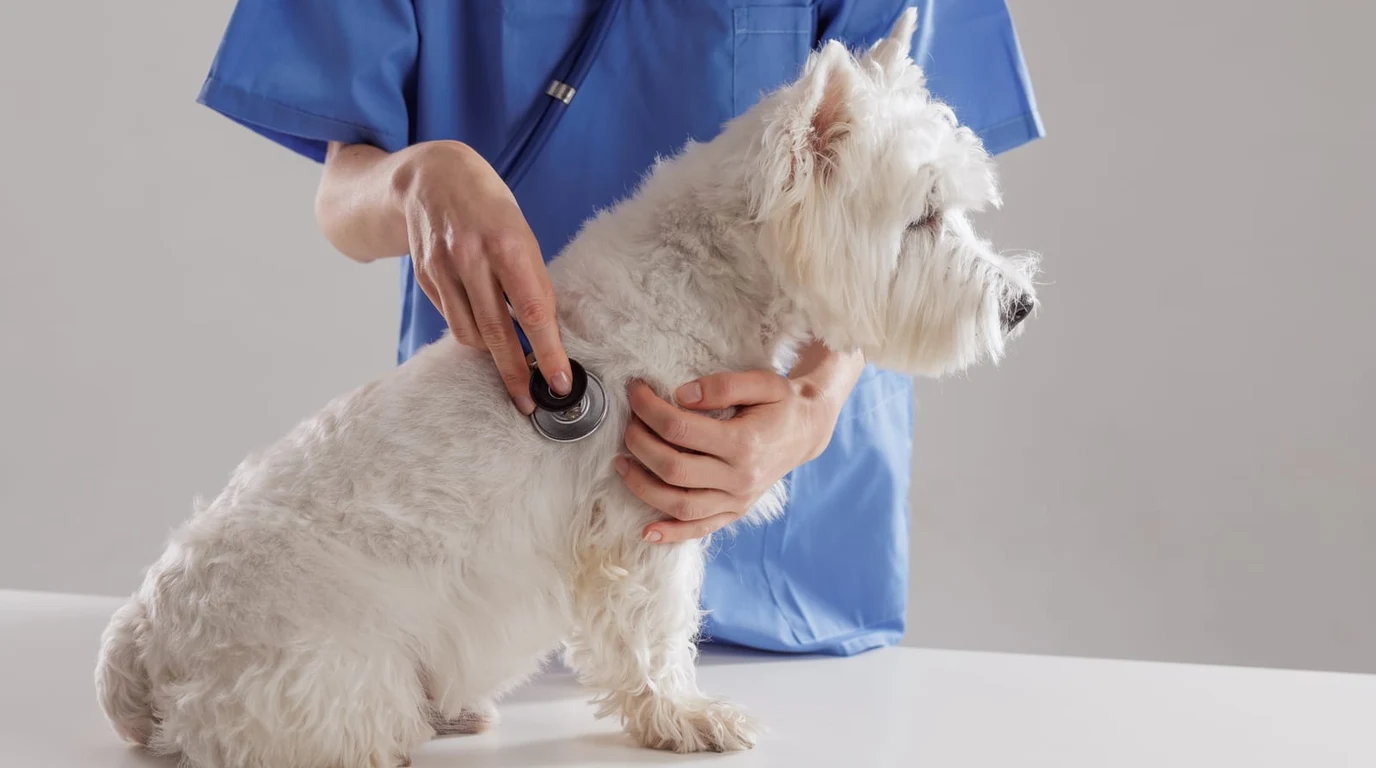
When an orthopedic veterinarian evaluates an acute or chronic hind limb lameness, diagnosing a CCL tear relies on precise manual physical examination techniques. Because ligaments are soft tissue structures that do not show up on plain radiographic x-rays, the diagnosis cannot be made by looking at standard bone films alone; rather, x-rays serve to assess secondary joint effusion (fat pad displacement), osteophyte formation along the patella and fabellae, and rule out bone tumors (osteosarcoma) or avulsion fractures.
The definitive physical examination maneuvers are the Cranial Drawer Test and the Tibial Compression Test (Cranial Tibial Thrust Test):
- The Cranial Drawer Test: With the dog standing or in lateral recumbency, the examiner stabilizes the distal femur with one hand and grasps the proximal tibia with the other. The examiner attempts to manually slide the tibia forward relative to the femur. In a joint with an intact cranial cruciate ligament, there is zero forward translation. In a complete CCL tear, the tibia shifts forward noticeably—resembling a drawer opening from a cabinet. In tense, muscular dogs or acute partial tears, voluntary muscle spasm can mask drawer movement, frequently necessitating examination under light sedation.
- The Tibial Compression Test: This test replicates the natural biomechanics of weight-bearing. The examiner flexes the hock (ankle) while holding the stifle in a fixed position. If the CCL is ruptured, flexing the hock causes the Achilles tendon mechanism to push the tibia forward automatically.
Multimodal Post-Operative Pain Protocols & Sedation
Modern veterinary surgery has evolved far beyond relying on a single post-operative pain medication. Contemporary TPLO surgical protocols employ an integrated multimodal analgesic strategy that blocks nociceptive pain pathways at the spinal, peripheral, and central nervous system levels:
- Targeted Peri-Operative Local Anesthesia: Advanced surgical centers administer a pre-operative femoral and sciatic nerve block (using bupivacaine or ropivacaine under ultrasound guidance). This completely numbs the entire hind limb for 12 to 24 hours post-surgery, significantly reducing the depth of general gas anesthesia required during the osteotomy and eliminating post-surgical emergence pain.
- NSAIDs (Non-Steroidal Anti-Inflammatories): Target cyclooxygenase-2 (COX-2) enzymes to suppress localized joint inflammation. Medications such as carprofen, meloxicam, or robenacoxib form the core foundation of pain relief for the initial 14 to 21 days, contingent on normal baseline renal and hepatic blood panels.
- Neuropathic Pain Modulation: Because cutting bone and joint capsule manipulation triggers neuropathic signaling, gabapentin is routinely prescribed to down-regulate central sensitization and alleviate bone healing discomfort.
- Confinement Sedatives (Trazodone): For young, high-drive, or anxious dogs (such as Australian Shepherds, Border Collies, or young Labradors), maintaining crate confinement for eight consecutive weeks is mentally challenging. Veterinary surgeons prescribe mild central serotonin antagonists (such as trazodone) to provide calm, stress-free relaxation, preventing high-energy surges that could fracture the healing bone.
Contralateral Knee Preservation: Protecting the Second Leg
Given the documented 40% to 60% probability of a dog tearing their opposite cruciate ligament within two years, the final stage of long-term orthopedic care must focus aggressively on contralateral limb preservation. The single most potent, scientifically validated protective intervention is strict canine weight optimization.
In canine biomechanics, every single kilogram of excess body fat multiplies the mechanical shear force exerted across the tibial plateau during trotting and galloping. Clinical trials demonstrate that reducing a dog’s Body Condition Score (BCS) from an overweight 7/9 down to a lean, athletic 4.5/9 reduces ground reaction loading by over 30%, significantly delaying or preventing the degradation of the remaining healthy cruciate fibers.
Furthermore, early initiation of targeted chondroprotective therapies—including intramuscular polysulfated glycosaminoglycan injections (Adequan), undenatured type II collagen (UC-II), and high-potency marine omega-3 fatty acids (EPA/DHA)—modulates matrix metalloproteinases in the uninjured joint, slowing cartilage degradation and preserving long-term mobility.
The 12-Week TPLO Rehabilitation & Recovery Roadmap
Recovering from TPLO surgery is a biological marathon that requires strict phased confinement, progressive physical therapy, and careful weight management. The standard 12-week rehabilitation timeline proceeds through four defined stages:
Stage 1: Acute Healing & Inflammation Control (Weeks 1 to 2)
- Confinement: 100% strict confinement to a small room, playpen, or crate with non-slip rugs. Absolutely zero running, jumping, stairs, or free roaming.
- Mobility: Leash walks strictly for elimination purposes only (maximum 3 to 5 minutes) using a support sling beneath the belly to prevent slipping.
- Cryotherapy & PROM: Apply cold packs wrapped in towels to the incision for 15 minutes three times daily. Perform gentle Passive Range of Motion (PROM) flexion and extension within the dog’s comfort zone.
Stage 2: Early Callus Formation & Controlled Weight-Bearing (Weeks 3 to 6)
- Incision Check: Surgical sutures/staples removed at day 10–14.
- Controlled Leash Walking: Gradually increase flat leash walks from 5 minutes to 10–15 minutes, two to three times daily. Keep pace slow to encourage the dog to place the foot flat with every stride.
- Targeted Proprioception: Introduce gentle weight-shifting exercises, controlled sit-to-stand repetitions (“dog squats”), and balance pad standing to rebuild quadriceps muscle mass.
Stage 3: Radiographic Union & Dynamic Strengthening (Weeks 7 to 8)
- 8-Week Recheck Radiographs: Mandatory orthogonal x-rays taken under light sedation by the veterinary surgeon to verify complete bone bridging (union) across the osteotomy site.
- Physical Therapy Progression: Once radiographic union is confirmed, the dog can begin advanced rehabilitation, including underwater treadmill hydrotherapy, stepping over cavaletti poles, and controlled uphill walking to restore hindquarter muscle symmetry.
Stage 4: Return to Full Functional Activity (Weeks 9 to 12)
- Off-Leash Conditioning: Leash walks extended to 30–45 minutes. Controlled, off-leash activity gradually reintroduced in enclosed, safe environments.
- Full Recovery Milestone: By week 12, over 90% of dogs achieve near-normal limb function, symmetry, and joint comfort, resuming active hikes, swimming, and canine sports.
Companion Joint & Recovery Masterclasses
Protecting orthopedic healthspan requires proactive biomechanical care. Explore our companion analyses on Canine Massage & Acupressure for Senior Joint Relief and Long Dog Nails and Postural Strain on Canine Knees.
People Also Ask
Can a dog live with a torn ACL without surgery?
Small dogs under 15 pounds with low activity levels can sometimes stabilize a torn CCL through conservative medical management, strict rest, and scar tissue formation. However, in dogs over 25 pounds, untreated CCL tears lead to severe joint instability, progressive meniscus tears, and debilitating, irreversible osteoarthritis.
How long after TPLO surgery can a dog walk?
Most dogs begin lightly “toe-touching” and bearing partial weight on the operated leg within 24 to 48 hours following TPLO surgery. Full weight-bearing typically occurs by week 2 to 3, but leash-only restriction is mandatory until bone healing is confirmed on x-rays at week 8.
What is the success rate of TPLO surgery in dogs?
Published veterinary orthopedic literature reports a 93% to 96% success rate for TPLO surgery, with the vast majority of dogs returning to full, active athletic performance with minimal long-term lameness.
Does pet insurance cover TPLO surgery in Canada?
Yes, provided the policy was in effect prior to the injury and the dog has passed the standard orthopedic waiting period (typically 6 months). However, if one knee tore prior to insurance enrollment, insurers classify both knees as pre-existing conditions and will deny coverage for the contralateral leg.