
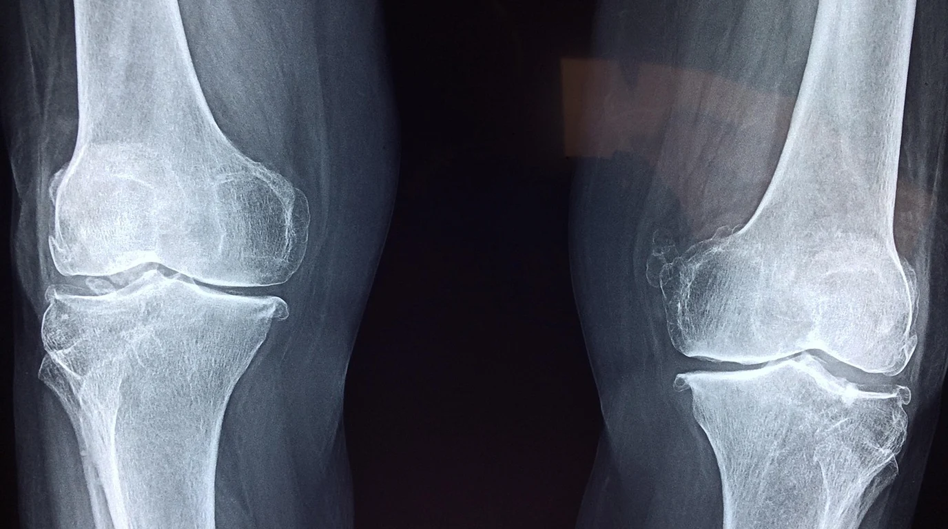
Hearing your dog emit a sudden, piercing yelp during an energetic game of fetch—followed by the immediate refusal to bear weight on a hind leg—is among the most gut-wrenching experiences a dog owner can face. In over 85% of acute canine hind-limb lameness cases, the diagnostic culprit is a rupture or partial tear of the Cranial Cruciate Ligament (CCL), the canine equivalent of the human Anterior Cruciate Ligament (ACL). However, unlike human athletes who tear their ACL through traumatic contact collisions, canine cruciate disease is almost universally a chronic, progressive degenerative arthropathy. The ligament fibers weaken over months due to biomechanical tibial slope angles, chronic systemic inflammation, and genetic conformation before final mechanical failure occurs. Faced with an acute tear, pet owners are thrust into a high-stakes surgical dilemma: orthopedic surgeons advocate for immediate, invasive Tibial Plateau Leveling Osteotomy (TPLO) surgery costing $4,500 to $7,000 per knee, while conservative management advocates tout custom carbon-fiber braces, regenerative PRP injections, and hydrotherapy. Here is Julian Hayes’s forensic biomechanical analysis of canine CCL tears, TPLO geometry vs conservative rehabilitation, long-term osteoarthritis progression, and how to determine the optimal recovery path for your dog.
Why Do Dogs Tear Their Cruciate Ligaments Differently Than Humans?
To visualize this biomechanical reality, imagine a wagon parked on a steep driveway. The cranial cruciate ligament acts as the rope preventing the wagon from rolling downhill. In humans, because our shin bone (tibia) is flat, our ACL only experiences tension during pivoting, jumping, or traumatic hyper-extension. In dogs, every single step taken throughout their entire lifetime exerts continuous shear tension on the CCL rope.
When the ligament begins to fray, the joint experiences micro-instability. The body attempts to stabilize the joint by producing fibrous scar tissue (the classic “medial buttress” thickening felt on the inner knee) and laying down osteophytes (bone spurs). If untreated, the sliding femur crushes the fibrocartilage medial meniscus, resulting in excruciating nerve pain, audible joint clicking, and accelerated degenerative joint disease.
Treatment Modality Comparison: TPLO vs Lateral Suture vs Conservative Rehab
To compare orthopedic intervention strategies across canine cruciate disease, evaluate this empirical breakdown:
| Treatment Modality | Surgical / Intervention Mechanics | Ideal Canine Candidate | Full Recovery Timeline | Long-Term Arthritis Resistance |
|---|---|---|---|---|
| TPLO Surgery | Radial osteotomy cuts tibia; rotates plateau to 5°; titanium plate fixation | Dogs >45 lbs; high-energy athletic dogs; steep TPA (>24°) | 8–12 Weeks (Bone union verified via X-ray) | Highest (Biomechanically neutralizes thrust permanently) |
| Extracapsular Lateral Suture | Heavy nylon mono-filament looped around fabella to mimic ligament | Small dogs <30 lbs; low-activity geriatric canines | 10–14 Weeks (Relies on fibrous scar formation) | Moderate (Suture stretches or snaps in heavy dogs) |
| Conservative Rehabilitation | Strict cage rest; custom dynamic orthotic brace; hydrotherapy; PRP | Small dogs (<25 lbs); anesthetic-risk seniors; financial constraints | 16–24 Weeks (Prolonged fibrosis stabilization) | Poor in large dogs (High risk of subsequent meniscal tear) |
The 50% Bilateral Tear Reality: Protecting the Opposite Knee
One of the most emotionally and financially exhausting realities of canine cruciate disease is the Bilateral Phenomenon. Veterinary orthopedic studies demonstrate that between 40% and 60% of dogs that tear one cranial cruciate ligament will tear the contralateral (opposite) ligament within 12 to 18 months.
Because the disease is degenerative rather than traumatic, both knees possess identical genetic geometry, collagen collagen degradation, and tibial plateau slope. When the first knee tears, the dog shifts 70% of its rear-end body weight onto the “good” leg during the subsequent weeks of lameness and surgical rehabilitation. This severe compensatory overload dramatically accelerates the failure of the remaining ligament. Protecting the opposite knee requires aggressive weight optimization (dropping 5% to 10% of excess body fat reduces stifle joint loading by 30%), daily joint chondroprotectants, and avoiding ballistic fetch games during rehabilitation.
Julian Hayes’s Step-by-Step CCL Recovery Playbook
Actionable Protocol for Cruciate Injury Management
- Obtain Sedated Orthopedic Radiographs and Drawer Test: Do not rely on visual gait analysis alone. A veterinarian must perform a Cranial Drawer and Tibial Compression test under mild sedation to assess stifle laxity and measure the exact Tibial Plateau Angle (TPA).
- Inspect Meniscal Integrity: If you hear an audible click or pop when your dog walks, the medial meniscus is likely torn. Conservative management will fail; torn meniscal cartilage acts like a pebble inside a shoe, causing continuous pain and demanding surgical debridement.
- Enforce Strict 8-Week Confinement Post-TPLO: The bone cut executed during TPLO requires rigid stability to achieve primary osteosynthesis. Use an indoor exercise pen, carpet runners over slippery hardwood floors, and a rear-support sling for bathroom breaks. Zero jumping or running for 8 weeks.
- Initiate Professional Underwater Treadmill Hydrotherapy: Beginning at Week 6 post-surgery (or Week 4 in conservative rehab), underwater treadmill therapy allows low-impact, buoyant muscle rebuilding without concussive impact on healing stifle joints.
Targeted Post-Operative Modalities: Class IV Laser and Targeted PEMF
Modern canine orthopedic recovery has moved far beyond simple cage rest. To accelerate osteotomy bone healing and control post-surgical capsular edema, veterinary rehabilitation specialists utilize Class IV Photobiomodulation (Cold Laser Therapy) and Targeted Pulsed Electromagnetic Field (tPEMF) Therapy (such as the Assisi Loop).
Class IV laser therapy delivers near-infrared photons (810nm and 980nm wavelengths) directly into the deep stifle joint tissue. Cytochrome c oxidase in mitochondrial membranes absorbs this light energy, stimulating adenosine triphosphate (ATP) synthesis, accelerating cellular fibroblast proliferation, and suppressing pro-inflammatory prostaglandins. Daily 15-minute tPEMF sessions stimulate microvascular nitric oxide production, reducing soft tissue pain and swelling by up to 50% while stimulating osteoblast activity along the radial bone cut.