
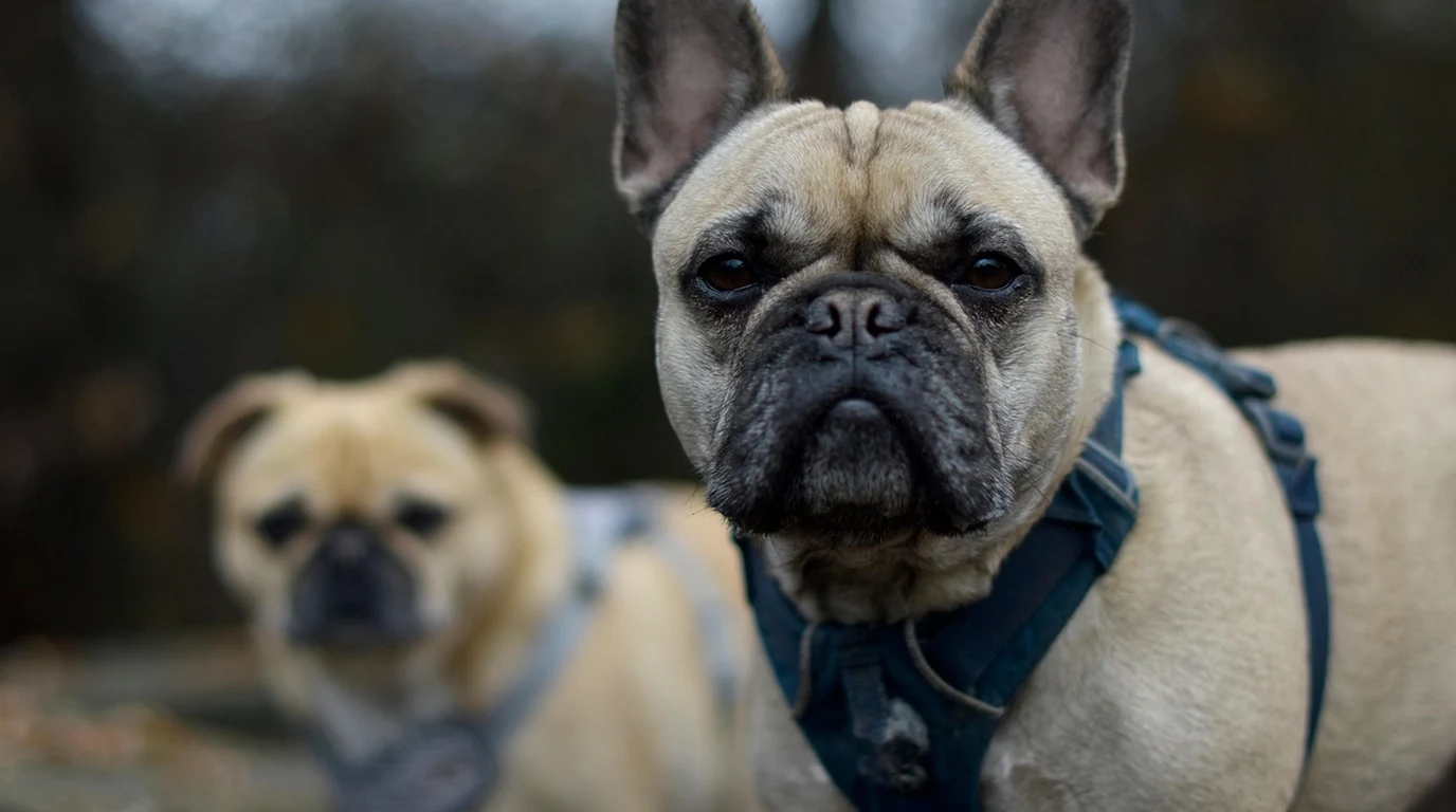
Few sounds in the canine world are as harrowing as the sudden, piercing scream of a French Bulldog suffering an acute intervertebral disc herniation. In a single horrifying second, a playful, energetic three-year-old Frenchie jumps off the family sofa, emits an agonized yelp, arches its back in rigid spasm, and collapses—its rear legs dragging limply across the floor like lifeless ropes. For decades, owners and breeders chalked up these devastating episodes to unpredictable bad luck or clumsy physical trauma. However, groundbreaking molecular genetics conducted at the UC Davis Veterinary Genetics Laboratory revealed the true underlying biological culprit: Chondrodystrophy (CDDY), driven by an aberrant FGF4 retrogene insertion on canine chromosome 12. This genetic mutation does not merely dictate short, bowed legs; it fundamentally corrupts the cellular biology of the canine spinal disc, turning pliant, shock-absorbing cartilage into brittle, calcified stone prone to catastrophic explosive extrusion. Here is Julian Hayes’s forensic veterinary guide to French Bulldog intervertebral disc disease (IVDD), understanding the difference between CDDY and CDPA genetic mutations, recognizing subtle early neurological warning signs, navigating the critical 24-hour surgical emergency window, and implementing proactive lifestyle architecture to protect your Frenchie’s spine.
How Does the CDDY Genetic Mutation Cause Disc Rupture in French Bulldogs?
To understand why French Bulldogs are uniquely vulnerable to spinal paralysis, one must examine the distinct genetics of canine dwarfism. In veterinary genetics, there are two distinct FGF4 retrogene insertions that produce short-legged phenotypes:
- Chondrodysplasia (CDPA — Chromosome 18): This ancient retrogene mutation is responsible for the classic short-limbed appearance of breeds like the Basset Hound and Dachshund. Crucially, CDPA alters long-bone growth plates (shortening the radius, ulna, and tibia) without triggering premature intervertebral disc degeneration.
- Chondrodystrophy (CDDY — Chromosome 12): This much more recent mutation acts as a semi-dominant trait for both leg shortening and intervertebral disc disease. Dogs carrying even a single copy of CDDY (N/CDDY) or two copies (CDDY/CDDY) experience accelerated degeneration of all 26 intervertebral discs throughout their spinal column.
In a genetically normal dog, the central core of an intervertebral disc—the nucleus pulposus—is composed of a gelatinous, water-rich matrix containing viable notochordal cells. This fluid core acts as a hydraulic shock absorber, evenly dispersing axial compression across adjacent vertebrae. In French Bulldogs carrying the CDDY mutation, notochordal cells die off within the first year of life. They are replaced by chondrocytes that deposit collagen and mineralize with calcium phosphate crystals. The disc transforms from a resilient hydraulic cushion into dry, brittle, unyielding chalk.
When the dog performs a routine physical movement—such as jumping down from a bed, twisting during play, or bounding down stairs—the outer fibrous ring (the annulus fibrosus) ruptures. The hardened, mineralized disc core violently explodes upward into the spinal canal (Hansen Type I IVDD), severely contusing and compressing the delicate descending tracts of the spinal cord.
Canine IVDD Neurological Severity Grading Matrix
Veterinary neurologists grade intervertebral disc herniation on an objective five-tier scale. Clinical prognosis and treatment pathways are strictly dictated by this neurological score:
| Neurological Grade | Clinical Presentation | Neurological Reflex Deficits | Recommended Clinical Path | Surgical Recovery Prognosis |
|---|---|---|---|---|
| Grade 1: Spinal Hyperesthesia | Severe spinal pain, arched back, muscle spasms; dog walks normally | Normal proprioception; zero motor or sensory deficits | Strict crate rest (6 weeks) + multimodal analgesia (gabapentin, NSAID) | 90%–95% conservative resolution |
| Grade 2: Ambulatory Paraparesis | “Drunken” wobbly gait; rear legs sway, cross over, or stumble | Delayed conscious proprioception (knuckling paws on floor) | Strict crate rest or early decompressive hemilaminectomy | 90%–95% surgical success |
| Grade 3: Non-Ambulatory Paraparesis | Dog cannot stand or walk on rear limbs; kicks or moves legs when supported | Absent proprioception; voluntary motor present but insufficient to stand | Emergency surgical referral for MRI/CT and decompression | 85%–90% surgical recovery |
| Grade 4: Paraplegia with Deep Pain | Complete paralysis of rear limbs; urinary retention or overflow incontinence | Zero voluntary motor; deep pain perception intact (dog reacts when toe bone pinched) | URGENT surgical emergency (Hemilaminectomy within 24 hours) | 80%–85% surgical recovery if operated promptly |
| Grade 5: Paraplegia Loss of Deep Pain | Complete rear paralysis; zero response when periosteum of toe pinched with hemostat | Complete sensory and motor obliteration; loss of nociceptive pathways | CRITICAL emergency surgery within 24 hours; risk of fatal myelomalacia | <50% recovery; drops to <10% if surgery delayed >48 hours |
Diagnostic Nuances: Why Plain X-Rays Cannot Diagnose Spinal Compression
A frequent and dangerous point of clinical friction occurs when a primary care veterinarian takes standard spinal radiographs (X-rays) and tells an anxious owner: “The spine looks clear, so it’s not a slipped disc.” This is a profound diagnostic misconception.
Plain radiographs visualize dense mineralized bone structures; they cannot visualize soft tissue neural parenchyma, nerve roots, or the spinal cord itself. While plain X-rays can occasionally reveal disc space narrowing or a calcified disc lying passively in the invertebral space, they cannot confirm whether a disc has extruded into the spinal canal or quantify the degree of spinal cord compression. Furthermore, dogs with severe spinal cord compression can have completely normal-appearing vertebral spacing on plain films.
The undisputed clinical gold standard for canine IVDD imaging is Magnetic Resonance Imaging (MRI), with Computed Tomography (CT) Myelography serving as an excellent secondary modality. MRI provides high-contrast cross-sectional images of the spinal cord parenchyma, revealing the exact anatomical location of disc herniation, distinguishing disc extrusion from fibrocartilaginous embolism (FCE) or spinal neoplasia, and identifying life-threatening progressive ascending/descending myelomalacia (spinal cord liquefaction necrosis).
Proactive Environmental Architecture: Protecting the Chondrodystrophic Spine
Because the CDDY mutation is virtually fixed in the French Bulldog gene pool, handlers must operate under the assumption that every Frenchie has brittle, calcified discs. You cannot alter their genetics, but you can systematically eliminate the mechanical forces that trigger disc rupture:
The Frenchie Spinal Preservation Protocol
- Enforce the Zero-Jumping Rule (Furniture Ramps): The single most common trigger for acute Hansen Type I disc rupture is landing from elevated furniture. The concussive impact of landing forces the spine into violent hyperextension and axial compression. Install shallow-incline, high-traction carpeted ramps beside beds and sofas, and systematically train your Frenchie to use them with positive reinforcement.
- Block Staircase Access: Navigating standard human staircases subjects the thoracolumbar junction (T11 to L3—the most frequent site of IVDD rupture) to continuous rotational shear stress. Install secure baby gates at both the top and bottom of all staircases. Carry your Frenchie up and down stairs using a level two-handed scoop technique that supports both the chest and rump horizontally.
- Eliminate Neck Collars (Harnesses Only): French Bulldogs have high rates of cervical disc disease (C2 to C5). Walking a Frenchie on a standard neck collar transfers leash tension directly into cervical vertebral discs. Walk your dog exclusively on a wide, padded Y-shaped chest harness that distributes force across the sternum.
- Maintain a Lean Body Condition Score (BCS 4.5/9): Excess adipose weight places continuous gravitational downward torque on the lumbar spine. Keeping your French Bulldog lean—where you can readily feel their ribs and observe a visible abdominal tuck—reduces spinal load by up to 30%.
- Recognize Early Subtle Pain Cries: French Bulldogs are stoic. Before paralysis occurs, they almost always display 24 to 48 hours of subtle discomfort: refusing to lower their head to eat from a floor bowl, shivering, hiding under furniture, or crying out when picked up under the armpits. If you notice these signs, initiate immediate crate rest and seek emergency veterinary evaluation.
Genetic predispositions require proactive multi-system monitoring. Compare oncological screening with orthopedic gene testing in our guide on Golden Retriever Cancer Prevention: Hemangiosarcoma.