
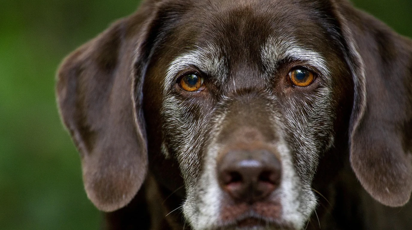
When our canine companions cross into their senior years—typically around age seven or eight for medium to large breeds—attentive dog guardians often notice a sudden, unsettling change in their dog’s eyes. Under bright indoor lighting or sunlight, the clear black pupils begin displaying a distinct, hazy, bluish-grey translucence. For many pet parents, this misty ocular clouding triggers immediate panic: fears of accelerating blindness, painful intraocular inflammation, and costly veterinary ophthalmic surgeries. However, in veterinary ophthalmology, the appearance of cloudy eyes in senior dogs bifurcates into two fundamentally distinct physiological phenomena: benign Nuclear Sclerosis (Lenticular Sclerosis) and pathological Cataracts. While cataracts represent a severe, progressive opacity of the lens that blocks light transmission and inevitably causes functional blindness, nuclear sclerosis is an entirely natural, pain-free aging phenomenon that preserves baseline functional vision throughout a dog’s natural lifespan. Distinguishing between these two conditions is essential to avoid unnecessary medical anxiety while ensuring true cataracts are identified and treated before secondary complications like lens-induced uveitis destroy the eye. Here is Julian Hayes’s clinical veterinary guide to nuclear sclerosis versus cataracts, slit-lamp ophthalmic examination protocols, tapetal reflection tests, and senior canine vision preservation.
What is the Biological Difference Between Nuclear Sclerosis and Cataracts?
To understand the optical divergence between these two conditions, one must examine the unique microscopic anatomy of the canine crystalline lens. The lens is an avascular, transparent biconvex structure responsible for focusing incoming light rays onto the sensory retina. Unlike epithelial skin cells or gastrointestinal lining cells that shed continuously, the canine lens never sheds a single cell throughout the dog’s life.
Throughout a dog’s life, the lens epithelium continuously produces new secondary lens fibers at the periphery (the cortex). As new fibers are laid down concentric to the capsule, older fibers are systematically pushed inward toward the central core (the nucleus). Over 8 to 12 years of continuous inward cellular compression, the nucleus becomes increasingly dense, dehydrated, and compact. This optical compaction changes the refractive index of the central lens, reflecting shorter blue wavelengths of light back toward the observer. Crucially, because the compressed fibers remain biochemically transparent, light continues traveling cleanly through the lens to the photoreceptor rods and cones of the retina. The dog experiences mild presbyopia (loss of close-up accommodation), but retains functional distance and navigational vision.
In stark contrast, a cataract occurs when lens protein structures (crystallins) undergo pathological denaturing, oxidative aggregation, and cellular breakdown. Disrupted metabolic balance—triggered by genetic mutations, diabetic sorbitol accumulation, blunt ocular trauma, or chronic inflammation—causes the normally transparent lens fibers to clump into milky, opaque white barriers. As cataracts mature, they physically block photons from reaching the retina, resulting in functional blindness.
Clinical Comparison: Nuclear Sclerosis vs Cataracts in Dogs
| Diagnostic Characteristic | Nuclear Sclerosis (Lenticular) | Canine Cataracts (Pathological) | Clinical Implication |
|---|---|---|---|
| Visual Appearance | Translucent bluish-grey pearlescent haze | Opaque milky white, marble, or cracked ice | Cataracts block internal ocular structures |
| Symmetry & Onset | Strictly bilateral and symmetric (Both eyes) | Often asymmetrical or unilateral initially | Asymmetric clouding strongly indicates cataracts |
| Tapetal Reflection (Eye Shine) | 100% Present (Green/Gold shine visible) | Blocked / Absent (Dark silhouette shadow) | Key diagnostic test using penlight |
| Functional Vision Impact | Minimal (Slight close-up focus loss) | Severe progressive blindness | Cataract dogs bump into furniture in dim light |
| Secondary Complications | Zero risk of uveitis or glaucoma | High risk: Lens-induced uveitis, glaucoma | Cataracts require anti-inflammatory drops |
| Treatment Protocol | None required (Benign physiological aging) | Phacoemulsification surgery + IOL implant | Surgery restores complete optical clarity |
The At-Home Flashlight Test: Evaluating the Tapetal Reflection
While an official diagnosis requires a veterinary ophthalmic examination using a direct ophthalmoscope or biomicroscopic slit-lamp following pupillary dilation (mydriasis), dog guardians can execute a reliable preliminary screening test at home using a standard penlight or smartphone flashlight:
- Dim Room Lighting: Bring your senior dog into a dimly lit room and allow their pupils to naturally dilate for three minutes.
- Position Flashlight at Eye Level: Hold the light source directly adjacent to your nose or eye line, pointing toward the dog’s face from a distance of four to six feet.
- Observe the Retinal Tapetal Reflection: Look for the bright greenish-yellow or golden “eye shine” (the tapetum lucidum reflection) reflected back from the retina.
- In Nuclear Sclerosis: The tapetal reflection shines brightly and uniformly through the lens, illuminating the entire pupil. Because the nuclear haze is transparent, the back of the eye remains fully visible.
- In Cataracts: The cataract acts as an optical barrier, casting a dark silhouette, partial shadow, or complete blackout over the tapetal reflection. If the reflection is obstructed or fragmented, a true cataract is present.
The Diabetic Cataract Emergency: Rapid Sorbitol Osmotic Swelling
A critical veterinary emergency occurs in dogs diagnosed with diabetes mellitus. In diabetic canines, chronic systemic hyperglycemia overwhelms the normal hexokinase metabolic pathway in the lens. Excess glucose is shunted into the sorbitol pathway, catalyzed by the enzyme aldose reductase.
Sorbitol cannot easily diffuse across the lens capsule, accumulating inside lens fibers and creating an extreme intracellular hyperosmotic gradient. Water from the aqueous humor rushes into the lens, causing massive cellular swelling, lens fiber rupture, and the sudden emergence of diabetic cataracts within 48 to 72 hours. These swelling lenses can develop radial cortical intumescence, pushing forward into the anterior chamber and triggering acute angle-closure glaucoma or lens capsule rupture. Any diabetic dog presenting with sudden ocular clouding requires immediate same-day emergency veterinary ophthalmology triage.
Nutritional & Antioxidant Interventions for Canine Eye Longevity
While no oral supplement can reverse established mature cataracts or dense nuclear sclerosis, clinical evidence demonstrates that targeted antioxidant nutrition can attenuate photo-oxidative stress and support senior ocular metabolic health:
- Lutein and Zeaxanthin (Macular Carotenoids): Dietary xanthophyll carotenoids that cross the blood-retinal barrier to accumulate in canine ocular tissues, quenching singlet oxygen radicals generated by ultraviolet light exposure.
- Astaxanthin: A marine microalgae-derived keto-carotenoid exhibiting exceptional free-radical scavenging capacity across both the lipid bilayer and aqueous humor.
- N-Acetylcarnosine (NAC) Eye Drops: Formulated as topical ophthalmic drops that penetrate the cornea into the anterior chamber, where carnosine acts as a chaperone peptide to mitigate early oxidative protein cross-linking in incipient cortical cataracts.
Surgical Intervention: When is Phacoemulsification Indicated for Canine Cataracts?
While nuclear sclerosis requires zero medical therapy, progressive cataracts that impair functional navigation demand surgical consultation before secondary complications destroy the ocular architecture. The gold-standard veterinary surgical procedure is Phacoemulsification with Intraocular Lens (IOL) Implantation.
Under general anesthesia and operating microscope magnification, a veterinary ophthalmic surgeon makes a 2.8mm corneal micro-incision. An ultrasonic probe vibrating at 40,000 Hz emulsifies and aspirates the denatured, cataractous lens proteins from inside the capsular bag. A folding acrylic artificial intraocular lens is then inserted, restoring 20/20 optical refraction.
Timing is critical: surgical success rates exceed 92% when phacoemulsification is performed while the cataract is in the immature to early mature stage. If owners delay surgery until the cataract becomes hypermature, the leaking lens proteins induce severe Lens-Induced Uveitis (LIU), secondary glaucoma, and posterior synechiae, drastically reducing surgical success and risking permanent ocular enucleation.