
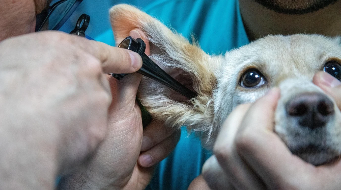
Few experiences in a dog guardian’s life are as gut-wrenching as running your hands under your companion’s jawline or behind their stifles and discovering firm, non-painful, golf-ball-sized swellings. Within hours at the veterinary clinic, a fine-needle aspirate (FNA) confirms every pet parent’s worst nightmare: canine lymphoma. Accounting for up to 24% of all canine cancer diagnoses, malignant lymphoma is one of the most common and aggressive hematopoietic neoplasms in companion animals.
Yet a diagnosis of canine lymphoma is not a monolithic death sentence. In modern veterinary oncology, lymphoma is not a single disease, but a diverse family of lymphoid malignancies with wildly divergent biological behaviors, chemotherapy responsiveness, and survival curves. The difference between an immunophenotype classified as B-cell lymphoma versus T-cell lymphoma represents the difference between a high-probability 12-to-24-month clinical remission and a refractory, drug-resistant disease course.
Navigating the complex landscape of staging diagnostics, flow cytometry, PARR genetic testing, multi-agent chemotherapy protocols (such as CHOP), and substantial veterinary financial costs requires objective scientific literacy. For devoted dog guardians, understanding the exact cellular math of lymphoma is the single most empowering step in making humane, compassionate, and life-extending decisions for their dog.
What Is the Difference Between B-Cell and T-Cell Canine Lymphoma?
Direct Answer: B-cell lymphoma originates from antibody-producing B lymphocytes (accounting for ~75% of cases) and responds exceptionally well to standard multi-agent CHOP chemotherapy, achieving 80–90% remission rates and 12–14 month median survival. T-cell lymphoma originates from thymus-derived T lymphocytes (~25% of cases), carries a poorer prognosis, and requires specialized alkylating protocols like MOPP or L-asparaginase.
In veterinary oncology training halls, the classic clinical mnemonic remains universal: “B is Better, T is Terrible.” While modern rescue protocols have significantly improved T-cell outcomes, this clinical aphorism accurately reflects the fundamental biological divergence between the two lineages. Lymphocytes are the primary white blood cells responsible for adaptive immunity. When clonal malignant transformation occurs, the cellular lineage dictates how the cancer cells interact with chemotherapeutic drugs.
B-cell lymphomas (most commonly Diffuse Large B-Cell Lymphoma, or DLBCL) typically present as multicentric lymphadenopathy—generalized swelling of external peripheral lymph nodes including submandibular, prescapular, axillary, inguinal, and popliteal nodes. Because B-cells retain active cell-surface receptors and predictable apoptotic pathways, they are remarkably sensitive to DNA-damaging agents. Conversely, T-cell lymphomas frequently present with hypercalcemia of malignancy (elevated blood calcium produced by parathyroid hormone-related peptide, PTHrP), mediastinal masses, and rapid intrinsic upregulation of P-glycoprotein multidrug resistance (MDR) efflux pumps that actively pump chemotherapy drugs out of tumor cells.
| Diagnostic Parameter | B-Cell Lymphoma (DLBCL) | T-Cell Lymphoma (Peripheral T-Cell) |
|---|---|---|
| Incidence Prevalence | ~70% to 80% of all multicentric cases | ~20% to 25% of cases |
| Primary Gold-Standard Chemo | 19-Week or 25-Week CHOP Protocol | MOPP, LOPP, or Tanovea (Rabacfosadine) |
| Initial Remission Rate | 85% to 90% Complete Remission (CR) | 50% to 65% (Often partial or short-lived) |
| Median Survival (Treated) | 12 to 14 months (20–25% reach 2+ years) | 6 to 8 months with standard protocols |
| Hypercalcemia Association | Rare (<5% of cases) | Frequent (Up to 40% present with high ionized calcium) |
| Untreated Survival Time | 4 to 6 weeks from onset | 3 to 5 weeks from onset |
The Critical Diagnostic Gate: Flow Cytometry vs. PARR
When a veterinarian performs an initial in-clinic fine-needle aspirate, cytology under a microscope can confirm lymphoma by identifying a monomorphic population of immature, intermediate-to-large lymphoblasts. However, standard cytology cannot distinguish between B-cell and T-cell lymphoma.
To determine immunophenotype and establish an accurate prognosis, two advanced laboratory diagnostics are employed:
1. Flow Cytometry
Flow cytometry is the preferred gold-standard test when live cellular samples can be rapidly shipped to a specialized veterinary reference laboratory (such as Colorado State University, North Carolina State, or IDEXX). Live cells extracted from a lymph node aspirate are suspended in specialized media and tagged with fluorescent antibodies targeting specific cell-surface clusters of differentiation (CD markers):
- CD21 and CD79a: Positive staining confirms B-cell lineage.
- CD3, CD4, and CD8: Positive staining confirms T-cell lineage.
- CD34: Indicates acute lymphoblastic leukemia or blast crisis, carrying significant negative prognostic implications.
2. PARR Testing (PCR for Antigen Receptor Rearrangements)
If live cells are unavailable, or if samples consist of archived formalin-fixed biopsy slides or dried cytology smears, PARR testing is utilized. PARR extracts genomic DNA and uses polymerase chain reaction (PCR) to evaluate the clonality of immunoglobulin (Ig) heavy chain genes (for B-cells) or T-cell receptor gamma (TCRγ) genes (for T-cells). While slightly less sensitive than flow cytometry, PARR is an exceptional confirmatory tool when cytology is ambiguous.
The single most devastating error in canine cancer management is starting prednisone before completing flow cytometry and definitive staging. When a general practice veterinarian sees swollen lymph nodes and prescribes prednisone to reduce swelling while the owner ‘thinks about chemotherapy,’ the steroid immediately induces apoptosis in malignant lymphoblasts. While this temporarily shrinks the nodes, it causes two catastrophic clinical consequences: first, it obliterates viable tumor cells, rendering flow cytometry inconclusive; second, and far more dangerous, prednisone rapidly upregulates the multidrug resistance gene (MDR1), inducing cross-resistance to doxorubicin and vincristine. Dogs exposed to prednisone prior to chemotherapy experience significantly shorter first remissions and higher rates of early relapse. Never administer prednisone until all staging biopsies and immunophenotyping are complete.
The World Health Organization (WHO) Canine Staging System
In addition to immunophenotype, veterinary oncologists classify lymphoma using the World Health Organization five-tier staging schema, supplemented by substage classification:
- Stage I: Involvement limited to a single solitary lymph node or tissue in a single organ (extremely rare).
- Stage II: Regional lymph node involvement on one side of the diaphragm (e.g., only submandibular and prescapular nodes).
- Stage III: Generalized lymph node enlargement involving nodes on both sides of the diaphragm (the most common clinical presentation).
- Stage IV: Liver and/or spleen enlargement (confirmed via abdominal ultrasound), with or without lymph node involvement.
- Stage V: Involvement of bone marrow (confirmed via marrow biopsy), blood, or non-lymphoid organs (central nervous system, eyes, skin).
Each stage is further subclassified into Substage a (clinically feeling well, normal appetite and energy, zero constitutional symptoms) or Substage b (systemically ill, exhibiting lethargy, anorexia, fever, weight loss, or vomiting). A dog in Stage IIIa carries a vastly superior statistical prognosis compared to a dog in Stage IIIb.
The CHOP Chemotherapy Protocol: Drugs, Cycles, and Tolerability
The undisputed cornerstone of canine lymphoma management is the cyclical multi-agent CHOP protocol (also known as the University of Wisconsin-Madison protocol). CHOP alternates four distinct antineoplastic drugs with non-overlapping mechanisms of action to prevent the emergence of drug-resistant cancer clones:
- C – Cyclophosphamide (Cytoxan): An alkylating agent that covalently binds to DNA, creating interstrand crosslinks that inhibit tumor cell replication. Administered orally or intravenously.
- H – Hydroxydaunorubicin (Doxorubicin / Adriamycin): An anthracycline antibiotic that intercalates DNA base pairs and inhibits topoisomerase II. Doxorubicin is the most potent single agent against canine lymphoma, administered as an intravenous infusion every three to four weeks.
- O – Oncovin (Vincristine): A vinca alkaloid derived from periwinkle plants that binds to tubulin proteins, halting cellular mitosis at metaphase. Administered via strict intravenous injection.
- P – Prednisone: An oral corticosteroid that binds to glucocorticoid receptors on malignant lymphoid cells, triggering rapid programmed cell death (apoptosis). Administered daily on a tapering scale.
| Treatment Modality | Typical Protocol Duration | Expected Median Survival | Estimated Veterinary Cost |
|---|---|---|---|
| Full 19-Week CHOP Protocol | 19 weeks (Weekly to bi-weekly visits) | 12 to 14 months (B-cell) | $6,500 – $10,000+ |
| Single-Agent Doxorubicin | 5 to 6 doses given every 3 weeks | 7 to 9 months | $2,500 – $4,500 |
| Oral CCNU (Lomustine) | Oral capsules given every 3 to 4 weeks | 4 to 6 months | $1,800 – $3,200 |
| Prednisone Alone (Palliative) | Daily oral medication until refractory | 2 to 3 months | $50 – $150 |
| No Treatment (Supportive Care) | Pain management / comfort care only | 4 to 6 weeks | Minimal |
The Reality of Canine Chemotherapy: Tolerability vs. Human Experience
The single greatest fear preventing pet guardians from pursuing chemotherapy is anthropomorphism: imagining their dog enduring the severe nausea, violent vomiting, total hair loss, and debilitating weakness often associated with human cancer treatment.
Veterinary oncology operates under a fundamentally different philosophical ethos than human medicine. In human oncology, the goal is often curative at all costs, pushing chemotherapy doses to the maximum tolerated threshold. In veterinary oncology, **the primary objective is clinical remission and outstanding quality of life**. Dosing protocols are calibrated to minimize toxicity:
- Over 80% to 85% of dogs experience zero to minimal side effects: Most dogs experience normal appetite, high energy, and complete happiness throughout their 19-week protocol.
- Fewer than 15% experience moderate GI upset: Managed rapidly with anti-nausea medications (maropitant/Cerenia, ondansetron) or metronidazole.
- Less than 5% require hospital supportive care: Rare instances of neutropenic fever or sterile hemorrhagic cystitis (from cyclophosphamide).
- Dogs do not lose their fur: Only continuous-growth breeds (Poodles, Bichon Frises, Schnauzers, and Old English Sheepdogs) experience thinning coat texture or whisker loss. Double-coated and short-haired breeds retain their full coats.
The 4:00 PM Actionable Playbook: Surviving a Lymphoma Diagnosis
- Immediate Fine-Needle Aspirate (FNA): If you detect swollen lymph nodes, insist on an immediate in-clinic aspirate. Confirm the presence of lymphoblasts cytologically.
- Order Flow Cytometry BEFORE Steroids: Explicitly instruct your veterinarian to submit an aspirate in saline/RPMI media for flow cytometry. Do not allow the administration of oral prednisone or steroid injections until this sample is safely drawn.
- Request a Board-Certified Oncology Consultation: General practitioners provide exceptional primary care, but specialized oncology centers possess access to clinical trials, precise CHOP dosing software, and rescue medications (such as Tanovea / rabacfosadine).
- Evaluate Quality-of-Life Metrics: Utilize the HHHHHMM Scale (Hurt, Hunger, Hydration, Hygiene, Happiness, Mobility, More Good Days Than Bad). Chemotherapy is only successful if your dog continues to experience joy, tail wags, and enthusiastic appetite.
🐾 Companion Guide: Senior Dog Clinical Bloodwork & Staging
Early detection of cellular changes requires annual baseline screening before external lymph nodes enlarge. Examine our comprehensive diagnostic audit:
👉 Read the Full Masterclass: Elevated Liver Enzymes in Dogs: ALT, ALP & Diagnosis »
People Also Ask
How long do dogs live with lymphoma without chemotherapy?
Without treatment, canine multicentric lymphoma progresses rapidly. The median survival time for untreated dogs is approximately four to six weeks from the onset of noticeable lymph node enlargement.
Can a dog be cured of lymphoma?
True permanent cures are extremely rare in canine oncology (occurring in under 2% to 5% of cases). The clinical goal of the CHOP protocol is prolonged, high-quality complete clinical remission where all detectable cancer disappears, returning the dog to vibrant health for 12 to 14 months or longer.
What is the relapse rate after CHOP chemotherapy?
Most dogs with B-cell lymphoma will eventually experience a relapse (typically between 9 and 12 months post-diagnosis). Upon relapse, re-induction with CHOP or transitioning to secondary rescue protocols (such as CCNU, MOPP, or Tanovea) achieves second remissions in approximately 60% to 70% of patients.
Is canine lymphoma painful?
In its early and intermediate stages (Stages I through IVa), multicentric lymphoma is generally painless. Swollen lymph nodes are non-tender to the touch. Pain typically arises only in end-stage disease involving bone marrow infiltration, secondary organ failure, or severe hypercalcemia.