
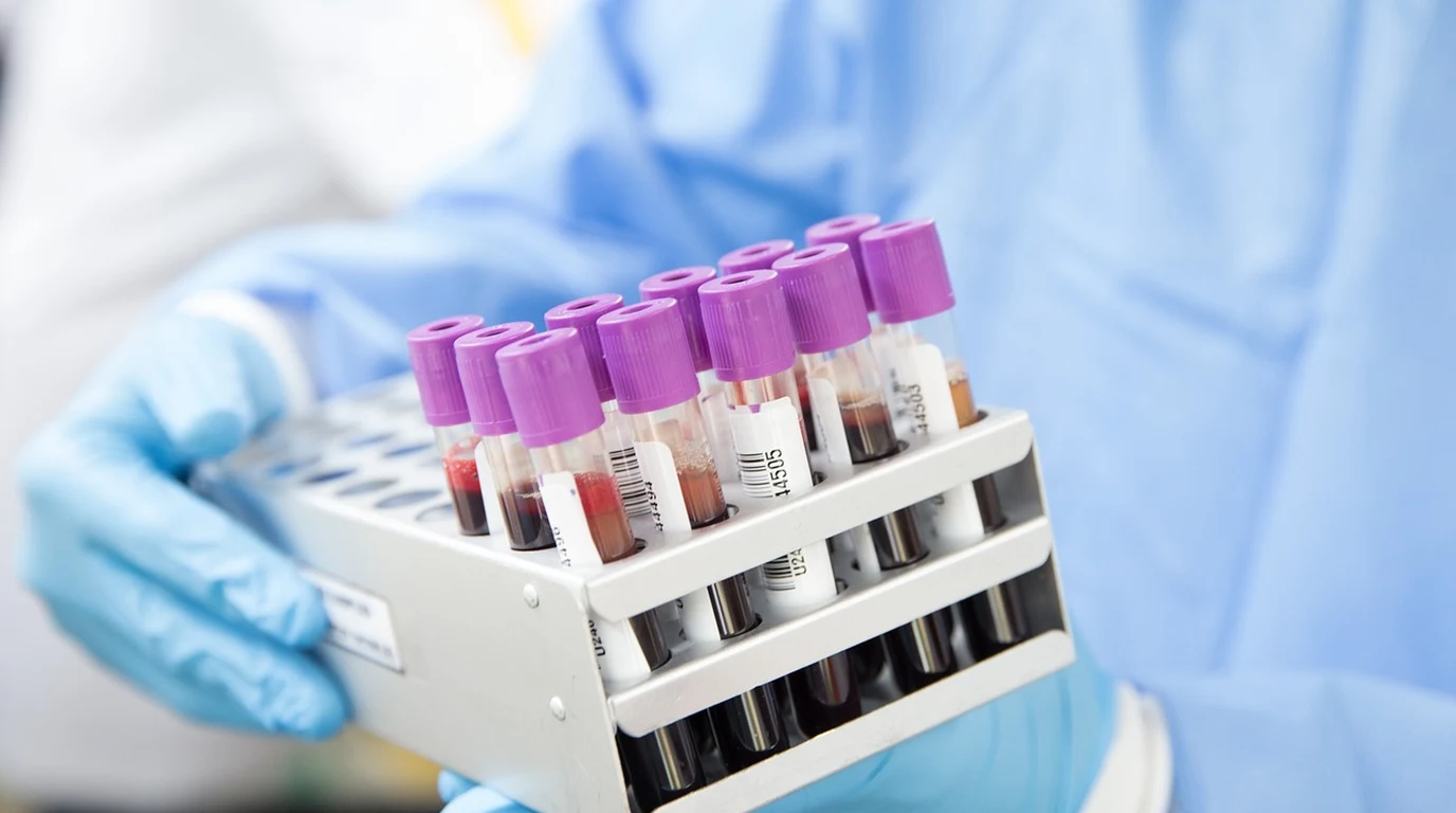
The phone rings, and it’s your vet. The words “liver enzymes are elevated” hit you like a cold wave. Immediately, your mind races. Is it serious? What does it mean for your beloved dog? You picture endless tests, specialist referrals, and the gnawing anxiety of not knowing. You’re not alone in that moment of dread; it’s a scenario played out in countless homes, sparking a frantic search for answers, for clarity, for a path forward that doesn’t feel like a bottomless pit of uncertainty and expense. This isn’t just about numbers on a lab report; it’s about the vibrant life force of your companion, and the deep, unspoken bond you share. You deserve more than medical jargon; you deserve a clear, actionable understanding.
### Are There Other Reasons My Dog’s ALP Could Be High Besides Liver Problems?
Absolutely, and this is perhaps the most critical piece of information for any dog owner facing an elevated ALP result. The biggest mistake is to immediately assume a primary liver disease when ALP is high. While liver involvement is certainly a possibility, ALP is a notoriously unspecific enzyme, and its elevation can be a red herring, pointing away from the liver rather than directly to it.
Let’s break down the common non-hepatic (non-liver) triggers for elevated ALP:
1. Corticosteroid Administration or Endogenous Overproduction (Cushing’s Disease): This is, without a doubt, the most frequent non-hepatic cause of elevated ALP in dogs.
* Exogenous Steroids: If your dog has been on prednisone, dexamethasone, or any other corticosteroid (even topical or inhaled forms can have systemic effects), their ALP will almost certainly be elevated, often significantly. This is due to the induction of the specific corticosteroid-induced ALP (C-ALP) isoenzyme in the liver, which is a normal physiological response, not a sign of liver damage.
* Cushing’s Disease (Hyperadrenocorticism): This endocrine disorder involves the overproduction of natural cortisol by the adrenal glands. The chronic excess of cortisol similarly induces C-ALP, leading to often dramatically high ALP levels. Other signs of Cushing’s include increased thirst and urination, increased appetite, pot-bellied appearance, thin skin, and hair loss. If these signs are present with high ALP, Cushing’s testing is warranted.
2. Bone Growth and Bone Disease:
* Young, Growing Dogs: Puppies and adolescent dogs naturally have higher ALP levels due to active bone growth and remodeling. This is completely normal and expected.
* Bone Tumors or Fractures: Any condition causing increased osteoblast activity (bone-forming cells), such as bone cancer (osteosarcoma) or healing fractures, can lead to elevated bone-specific ALP (B-ALP).
3. Gallbladder Mucocele: While technically affecting the biliary system (which is part of the liver’s plumbing), a gallbladder mucocele is a distinct condition where thick, gel-like mucus accumulates in the gallbladder, potentially obstructing bile flow. This obstruction leads to cholestasis and a subsequent rise in liver-specific ALP (L-ALP). It’s a serious condition that can lead to gallbladder rupture.
4. Other Systemic Illnesses: Sometimes, severe illness or inflammation elsewhere in the body can cause a mild, non-specific elevation in ALP, though typically not to the dramatic levels seen with steroids or Cushing’s. This is often a stress response.
5. Certain Medications (Non-Steroidal): While less common than corticosteroids, some other drugs can occasionally cause mild ALP elevations. It’s always crucial to inform your vet of all medications and supplements your dog is receiving.
Understanding these non-hepatic influences is paramount. Jumping to conclusions about liver disease based solely on an elevated ALP can lead to unnecessary anxiety, expensive and invasive procedures like liver biopsies, and, most importantly, a missed diagnosis of the true underlying condition, such as Cushing’s disease, which requires its own specific management. This is the essence of the “costly trap” – misdirection due to incomplete understanding.
### What’s the Smartest Next Step When My Dog Has High Liver Enzymes?
Navigating elevated liver enzymes requires a methodical, evidence-based approach. This isn’t a one-size-fits-all problem, and the “gold-standard diagnostic cascade” is designed to systematically rule out possibilities and pinpoint the true cause without unnecessary detours. Here’s your actionable playbook:
1. Re-evaluate and Re-test (The Initial Pause):
* Context is King: Before anything else, your vet should thoroughly review your dog’s history. Has your dog been on any medications (especially steroids)? Any recent stress, dietary changes, or exposure to toxins? Is your dog showing any clinical signs (lethargy, vomiting, diarrhea, jaundice, increased thirst/urination, appetite changes)?
* Fasting Blood Sample: Ensure the initial blood draw was from a fasted dog (typically 8-12 hours without food, water is fine). Post-prandial (after eating) samples can sometimes cause transient, mild ALP elevations.
* Repeat Bloodwork: Often, the first step is to repeat the blood test, especially if the elevation is mild, your dog is asymptomatic, or there’s a possibility of a transient factor. Sometimes, levels normalize on their own, indicating a temporary stressor or minor insult. This simple step can save significant worry and expense.
2. The Fasting/Post-Prandial Serum Bile Acids Test (The Liver Function Check):
* Why it’s crucial: While ALT and ALP indicate liver damage or stress, they don’t directly assess how well the liver is actually functioning. The serum bile acids test is the gold standard for evaluating liver function. Bile acids are produced by the liver, stored in the gallbladder, released to aid digestion, and then reabsorbed by the liver.
* How it works: Blood samples are taken after a 12-hour fast, then again 2 hours after a small, fatty meal. The liver’s ability to clear bile acids from the blood is measured.
* Interpretation: If both fasting and post-prandial bile acid levels are normal, it strongly suggests that despite elevated enzymes, the liver’s overall function is good, and the prognosis is often favorable. If bile acids are elevated, it indicates significant liver dysfunction, portal systemic shunting, or severe cholestasis, necessitating further investigation. This test helps differentiate between incidental enzyme elevations and true functional liver disease.
3. Abdominal Ultrasound (The Visual Inspection):
* What it offers: If bile acids are elevated, or if clinical signs warrant, an abdominal ultrasound provides a non-invasive visual assessment of the liver, gallbladder, and surrounding organs.
* What it can detect:
* Liver size and architecture: Enlargement, masses, abnormal texture.
* Gallbladder issues: Mucoceles, stones, inflammation (cholecystitis).
* Bile duct obstruction: Dilation of bile ducts.
* Other organ involvement: Pancreatitis, adrenal gland abnormalities (relevant for Cushing’s).
* Expertise matters: This procedure is best performed by an experienced veterinarian or a board-certified veterinary radiologist for the most accurate interpretation.
4. Liver Aspirate / Biopsy (The Definitive Diagnosis):
* When it’s needed: If the previous steps indicate significant, persistent liver disease, or if a mass is identified, a liver aspirate (fine needle aspiration) or biopsy may be recommended.
* Aspirate: A less invasive procedure where a small needle collects cells for cytological examination. It’s good for identifying certain types of inflammation, infection, or some cancers. However, it provides limited architectural information.
* Biopsy: A more invasive procedure (either surgical or ultrasound-guided) that obtains a tissue sample for histopathological examination. This is the gold standard for definitive diagnosis, allowing pathologists to assess cellular architecture, identify specific types of inflammation, fibrosis, necrosis, or cancer, and determine the severity and prognosis.
* Risks: Both procedures carry risks, including bleeding, infection, and potential for complications, especially in animals with compromised liver function (which can affect clotting). These risks must be carefully weighed against the benefits of a definitive diagnosis.
This cascade ensures that you’re not chasing shadows. You start with the least invasive, most informative tests and only progress to more invasive options when absolutely necessary, guided by the results of each preceding step.
### How Can I Support My Dog’s Liver Health Based on Evidence?
Once a diagnosis is reached, or while awaiting definitive results, your veterinarian may recommend supportive therapies. It’s crucial that these are evidence-based and tailored to your dog’s specific condition. Generic “liver support” supplements can be ineffective or, in some cases, even harmful if not chosen wisely.
Here are two of the most well-researched and commonly recommended hepatic support agents:
1. S-Adenosylmethionine (SAMe): The Master Antioxidant Precursor
* What it is: SAMe is a naturally occurring molecule found in every cell of the body. It plays a critical role in numerous biochemical reactions, including methylation, transsulfuration, and aminopropylation pathways.
* How it supports the liver:
* Glutathione Production: SAMe is a precursor to glutathione, often called the “master antioxidant” of the body. Glutathione directly neutralizes harmful free radicals, protecting liver cells from oxidative damage, which is a common feature of liver disease.
* Cell Membrane Fluidity: It helps maintain the integrity and fluidity of liver cell membranes, which are crucial for normal liver function.
* Detoxification: By supporting glutathione levels, SAMe enhances the liver’s natural detoxification processes.
* Evidence: Numerous studies in both humans and animals support SAMe’s hepatoprotective effects, particularly in cases of acute and chronic liver injury.
* Administration: SAMe supplements are typically given on an empty stomach, as food can significantly reduce its absorption. It’s available in various veterinary formulations.
2. Silybin (from Milk Thistle): The Anti-Inflammatory & Antioxidant Powerhouse
* What it is: Silybin is the most active component of silymarin, an extract from the milk thistle plant (Silybum marianum).
* How it supports the liver:
* Antioxidant Activity: Like SAMe, silybin is a potent antioxidant, scavenging free radicals and reducing oxidative stress in liver cells.
* Anti-inflammatory Effects: It helps to modulate inflammatory pathways, reducing the inflammatory response within the liver.
* Anti-fibrotic Properties: Some research suggests silybin may help inhibit the development of liver fibrosis (scarring), a serious consequence of chronic liver disease.
* Cell Regeneration: It may stimulate protein synthesis, potentially aiding in the regeneration of damaged liver cells.
* Evidence: Milk thistle and silybin have been extensively studied for their hepatoprotective properties, showing benefits in various forms of liver disease.
* Administration: Silybin is often combined with phosphatidylcholine (a phospholipid) to enhance its bioavailability, as silybin alone is poorly absorbed. It can be given with or without food, depending on the specific formulation.
Beyond Supplements: Addressing the Root Cause
It’s critical to remember that SAMe and silybin are supportive therapies. They help the liver cope and recover, but they do not cure the underlying disease. The most effective “actionable playbook” always involves:
* Treating the Primary Condition: If Cushing’s disease is diagnosed, specific medications (e.g., trilostane) are needed. If a gallbladder mucocele is present, surgical removal might be necessary. If a specific toxin or medication is identified, it must be removed or discontinued.
* Dietary Management: A low-fat, highly digestible diet may be recommended, especially for dogs with cholestasis or pancreatitis. Specific therapeutic diets formulated for liver disease can help reduce the metabolic burden on the liver.
* Fluid Therapy & Electrolyte Balance: For acutely ill dogs, intravenous fluids and correction of electrolyte imbalances are crucial.
* Antibiotics: If a bacterial infection is suspected or confirmed (e.g., cholangiohepatitis), appropriate antibiotics are essential.
* Anti-emetics & Appetite Stimulants: If your dog is nauseous or refusing to eat, medications to alleviate these symptoms are vital to prevent further deterioration.
Your role as a proactive owner, armed with this knowledge, is to work closely with your veterinarian. Ask questions, understand the rationale behind each diagnostic step and treatment recommendation, and advocate for your dog’s best interests. Elevated liver enzymes are a signal, not a sentence. With a thoughtful, systematic approach, many dogs with elevated ALT and ALP can go on to live full, healthy lives.
### Frequently Asked Questions
### What does it mean if my dog’s ALT is very high but ALP is normal?
If your dog’s ALT is significantly elevated while ALP remains normal, it strongly suggests acute hepatocellular damage or inflammation without significant cholestasis (bile flow obstruction) or steroid induction. This pattern points directly to primary liver cell injury, potentially from toxins, infections, or acute hepatitis, and warrants prompt investigation into the cause of the acute cellular insult.
### Can stress or diet cause elevated liver enzymes in dogs?
While severe stress or a sudden, drastic dietary change can sometimes cause mild, transient elevations in liver enzymes, especially ALP, it’s generally not a primary cause of significant or persistent elevations. A truly high ALT or ALP usually indicates a more specific underlying issue. However, ensuring a fasted blood sample and ruling out recent dietary indiscretions is always a good initial step.
### How long does it take for liver enzymes to return to normal after treatment?
The time it takes for liver enzymes to normalize depends entirely on the underlying cause, the severity of the damage, and the effectiveness of treatment. ALT, with its shorter half-life, may decrease within days to weeks once the acute damage stops. ALP, especially if steroid-induced, can take weeks to months to return to normal due to its longer half-life and the time required for the body to clear the inducing agent. Regular re-testing guides the assessment of treatment efficacy.
### Is elevated ALP always a sign of Cushing’s disease in older dogs?
No, elevated ALP in older dogs is not always a sign of Cushing’s disease, but it is a very common and important differential diagnosis to consider, especially if other clinical signs of Cushing’s are present (increased thirst/urination, pot-belly, hair loss). Other causes like gallbladder issues, certain medications, or even some non-hepatic tumors can also elevate ALP in older dogs. A thorough diagnostic workup is essential to differentiate.
Direct Practical Verdict:
### Why Did My Vet Say My Dog’s Liver Enzymes Are High?
When your veterinarian mentions “elevated liver enzymes,” they’re typically referring to two key players on your dog’s blood chemistry panel: Alanine Aminotransferase (ALT) and Alkaline Phosphatase (ALP). These aren’t actually enzymes that cause liver damage; rather, they are indicators, like smoke detectors, signaling that something might be amiss within the liver or, in ALP’s case, elsewhere in the body. Understanding their distinct roles is the first critical step in navigating this often-confusing diagnosis.
Think of your dog’s liver as a bustling, multi-tasking factory, essential for digestion, detoxification, nutrient storage, and protein synthesis. Within the liver’s cells, called hepatocytes, and along its intricate bile ducts, these enzymes reside. When these cells are damaged or stressed, or when certain physiological processes are activated, these enzymes can leak into the bloodstream or be produced in higher quantities, leading to their elevated levels on a blood test.
The initial concern is natural. High enzyme levels can point to serious liver disease, from acute hepatitis to chronic conditions, or even liver cancer. However, the magnitude of the elevation, the specific enzyme involved, and the presence of other clinical signs are all crucial pieces of the puzzle. A mild, transient elevation might be insignificant, while a dramatic, persistent rise demands immediate, thorough investigation. The trap many owners, and sometimes even practitioners, fall into is reacting solely to the numbers without considering the full clinical picture. This can lead to premature panic, unnecessary expensive tests, or, conversely, a dangerous delay in addressing a serious underlying issue. Our goal here is to equip you with the knowledge to discern the signal from the noise, to ask the right questions, and to collaborate effectively with your veterinary team.
### What’s the Real Difference Between Elevated ALT and ALP in My Dog’s Blood Work?
This is where the nuance truly begins, and it’s a distinction often oversimplified. ALT and ALP, while both “liver enzymes,” tell fundamentally different stories about what’s happening inside your dog. Misinterpreting their individual messages is a common and costly trap.
Alanine Aminotransferase (ALT): The Hepatocyte Damage Alarm
ALT is primarily found in the cytoplasm of hepatocytes, the main functional cells of the liver. It’s a “leakage enzyme,” meaning it’s normally contained within these cells. When hepatocytes are damaged, inflamed, or dying, their cell membranes become compromised, and ALT leaks out into the bloodstream.
* What it indicates: An elevated ALT is a strong indicator of acute hepatocellular damage or inflammation. Think of it as a direct alarm bell for the liver cells themselves. The higher the ALT, generally, the more significant the acute damage.
* Causes of elevation:
* Acute hepatitis: Inflammation of the liver, often due to toxins (e.g., certain medications, mushrooms, xylitol), infections (bacterial, viral, leptospirosis), or autoimmune conditions.
* Hypoxia: Lack of oxygen to liver cells, such as during severe shock or heart failure.
* Trauma: Direct injury to the liver.
* Certain medications: Some drugs, even those prescribed for other conditions, can be hepatotoxic (damaging to the liver).
* Severe muscle damage: While primarily liver-specific, very high levels of muscle damage can sometimes cause a mild ALT elevation, but this is less common.
* Key characteristic: ALT has a relatively short half-life in the bloodstream (around 2-3 days in dogs), meaning its levels rise quickly with acute damage and should decline relatively quickly once the damage ceases. Persistent elevation suggests ongoing injury.
Alkaline Phosphatase (ALP): The Inducible Enzyme with Many Hats
ALP is a more complex enzyme because it exists in several different forms, or isoenzymes, and is found in various tissues throughout the body, not just the liver. It’s an “inducible enzyme,” meaning its production can be ramped up in response to certain stimuli.
* What it indicates: Elevated ALP can signal cholestasis (impaired bile flow) within the liver, but it can also be elevated due to non-hepatic causes. This dual nature is precisely why ALP elevations are so often misinterpreted.
* Liver-specific ALP (L-ALP): This isoenzyme is located on the cell membranes lining the bile canaliculi (tiny ducts that transport bile). When bile flow is obstructed (cholestasis), these cells are stimulated to produce more L-ALP, which then leaks into the bloodstream.
* Causes of cholestasis: Gallbladder mucocele, bile duct obstruction (e.g., by a stone, tumor, or inflammation), severe diffuse liver disease.
* Corticosteroid-induced ALP (C-ALP): This is a unique isoenzyme in dogs that is produced in response to glucocorticoids (steroids).
* Causes of C-ALP elevation: Administration of exogenous corticosteroids (e.g., prednisone, dexamethasone), or endogenous overproduction of cortisol due to Cushing’s disease (hyperadrenocorticism). This is a very common cause of elevated ALP in dogs and often has nothing to do with primary liver disease.
* Bone ALP (B-ALP): Found in osteoblasts (bone-forming cells).
* Causes of B-ALP elevation: Rapid bone growth in young, growing dogs, bone tumors, or bone healing after fractures.
* Key characteristic: ALP has a longer half-life than ALT (around 3 days for L-ALP, longer for C-ALP), so its levels can remain elevated for longer periods, even after the initial stimulus has resolved.
The “costly trap” here is assuming any ALP elevation automatically means severe liver disease. While significant ALP elevation can indicate serious liver issues like cholestasis or severe diffuse liver damage, it’s equally, if not more, likely to be influenced by non-hepatic factors, especially in older dogs or those on certain medications. Overlooking these non-hepatic causes can lead to unnecessary, invasive, and expensive liver biopsies when the real problem lies elsewhere.
Elevated ALT primarily signals acute liver cell damage, while elevated ALP often indicates bile flow issues or non-hepatic factors like steroid use, bone growth, or Cushing’s disease. Interpreting these requires a holistic view, considering clinical signs, medication history, and further diagnostics like bile acids tests or ultrasound to pinpoint the exact cause and guide appropriate, targeted treatment, avoiding unnecessary interventions.
| Enzyme | Primary Location & Type | Key Clinical Significance | Common Non-Hepatic Influences | Typical Half-Life (Dogs) |
|---|---|---|---|---|
| Alanine Aminotransferase (ALT) | Hepatocyte cytoplasm; Leakage enzyme | Acute hepatocellular damage or inflammation. Direct indicator of liver cell injury. | Severe muscle damage (rarely significant elevation). | ~2-3 days |
| Alkaline Phosphatase (ALP) | Cell membranes (bile canaliculi, bone, steroid receptors); Inducible enzyme | Cholestasis (impaired bile flow), steroid induction, bone growth/turnover. | Corticosteroid administration (oral/injectable), Cushing’s disease, bone growth (young dogs), bone tumors, some non-hepatic tumors. | ~3 days (L-ALP), longer for C-ALP |
Veterinary Blood Biomarkers & Organ Longevity Diagnostics
Metabolic organ panels require early diagnostic interpretation to preserve functional nephrons and hepatocyte capacity. For liver biomarker analysis, review Canine Kidney Disease Stages: IRIS Guidelines 2026.